The Institute's activity in the field of computer vision was initiated by the Termowet measurement system - an award-winning device for automatic measurements of the surface properties of metals and their alloys, carried out at the interface of the liquid and solid phases. The system performs measurements at temperatures up to 1700° C. Using dedicated image processing and analysis algorithms developed at the Institute, the system determines the exact values of contact angles and surface tension, based on images of drops of liquid metals or other tested materials.
Since then, other computer-aided diagnostic tools have been developed for materials science, biology, food processing and the medical industry, based mainly on optical coherence tomography/X-ray tomography and MRI images.
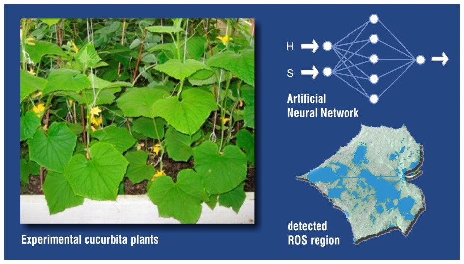
Recognition of reactive areas of oxygen compounds (ROS) in leaves using artificial neural networks [1].
Measurement of plant tissue activity and assessment of changes in root systems in response to biotic and abiotic factors.
An important area of the Institute's activity is also the development of image processing and analysis algorithms dedicated to medical imaging diagnostic support systems. Over the years, the Institute has developed many solutions to support doctors in their daily work. They include, among others:
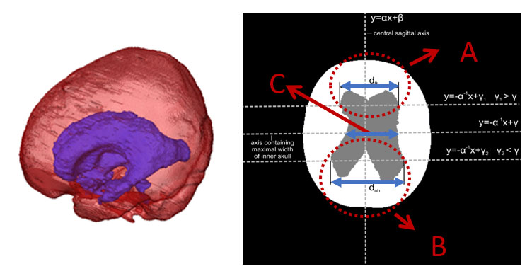
- assessment of neurological changes in the brain in children, in particular hydrocephalus and cranial deformities (cranostenosis) [2](*);
- ongoing monitoring of lung aeration changes in patients with CODP and ARDS [3];
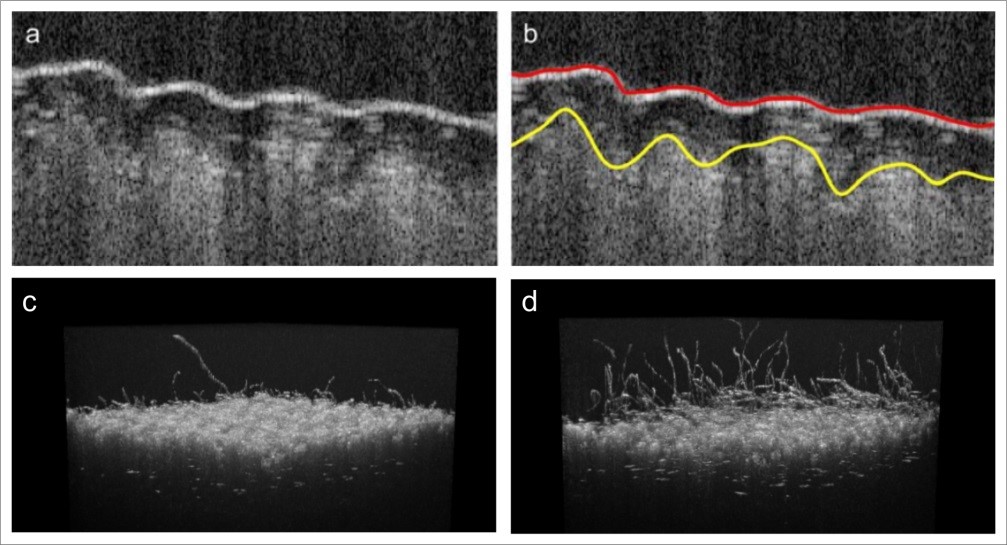
- assessment of the health of the corneal endothelium [4];
- analysis of contrast change curves for the purposes of supporting imaging diagnostics of prostate cancer [5]
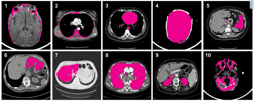
The Institute also develops general-purpose algorithms dedicated to improving the quality, skeletonization and segmentation of medical images.
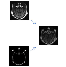
- Overlaying images using the wavelet transform [6].
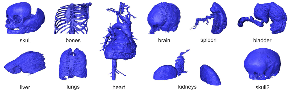
- Segmentation of three-dimensional medical images using the modified Random Walker method [7]
In particular, it concerns tomographic images of various modalities - optical coherence tomography (OCT) and X-ray tomography images.
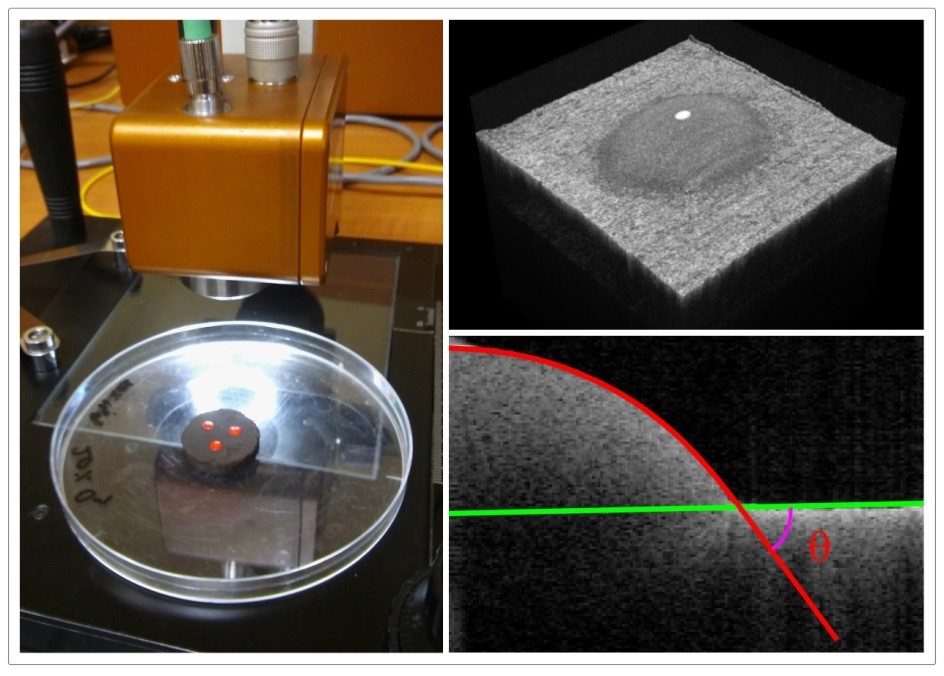
OCT image processing (**): Evaluation of the wettability of chemically modified lignite to determine its susceptibility to biosolubilization. Left: Spark OCT-1300 scanning head, right: 3D OCT image of a sessile droplet on a tablet made of compressed carbon powder; cross-section of a drop with a marked contact angle at the junction of three phases.
Evaluation of textile properties using images from optical coherence tomography (OCT) [8].
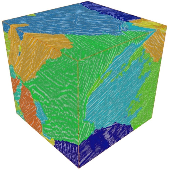
Detection of structural changes in X-ray tomographic images of selected materials (titanium alloys, bulk material) - Methods of volumetric image processing
- 3D texture orientation segmentation in the lamellar microstructure of a titanium alloy [9]
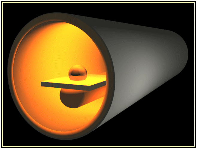
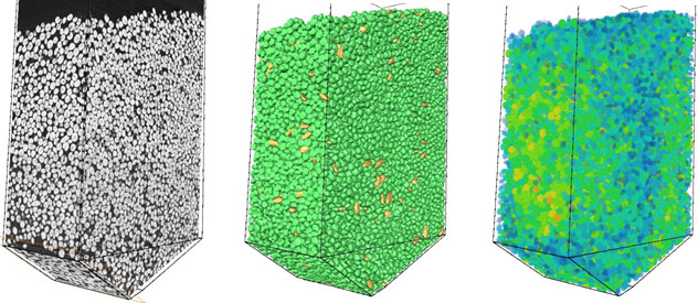
- Watershed segmentation, shape classification and extraction of physical properties of bulk materials during gravity flow in silos [10](***)
[1] J. Sekulska-Nalewajko, et al. Methods, 2016. 109: 114-133. https://doi.org/10.1016/j.ymeth.2016.05.018
[2] A. Fabijańska, T. Węgliński, et al. Int. J. Ap. Math. Comp., 2014, 24:299-312. http://dx.doi.org/10.2478/amcs-2014-0022
[3] D. A. Gómez Betancur, A. Fabijańska, et al. LNCS, 2016, 9972:395-407. https://doi.org/10.1007/978-3-319-46418-3_35
[4] A. Fabijańska. Artif. Intell. Med., 2018. 88: 1-13. https://doi.org/10.1016/j.artmed.2018.04.004
[5] A. Fabijańska. Comput. Biol. Med., 2016, 73:119-130. https://doi.org/10.1016/j.compbiomed.2016.04.010
[6] A. Bengueddoudj, Z. Messali Z, V. Mosorov. J. Innov. Opt. Heal. Sci., 2017. 10: 1750001. https://doi.org/10.1142/S1793545817500018
[7] A. Fabijańska, J. Gocławski. IEEE Im. Proc., 2015. 24: 524-537. https://doi.org/10.1109/TIP.2014.2383323
[8] J. Gocławski, et al. Polymers, 2018. 10:469. https://doi.org/10.3390/polym10050469
[9] R. Al Darwich, L. Babout. IET Im. Proc., 2018. 12: 1265-1272. https://doi.org/10.1049/iet-ipr.2016.0842
[10] L. Babout, K. Grudzień, et al. Compag, 2020. 172: 105346. https://doi.org/10.1016/j.compag.2020.105346
(*) Juventus Plus (IP2012 011272), A. Fabijańska (coordinator), J. Gocławski, T. Węgliński,E. Nowosławska, K. Zakrzewski, 2013-2015
(**) Large equipment grant MNiSW (6224/IA/138/2012), J. Sekulska-Nalewajko (coordinator), 2012
(***) Grant NCN OPUS10 (2015/19/B/ST8/02773), L. Babout (coordinator), K. Grudzień, S. Waktola, K. Miśkiewicz, 2016-2018